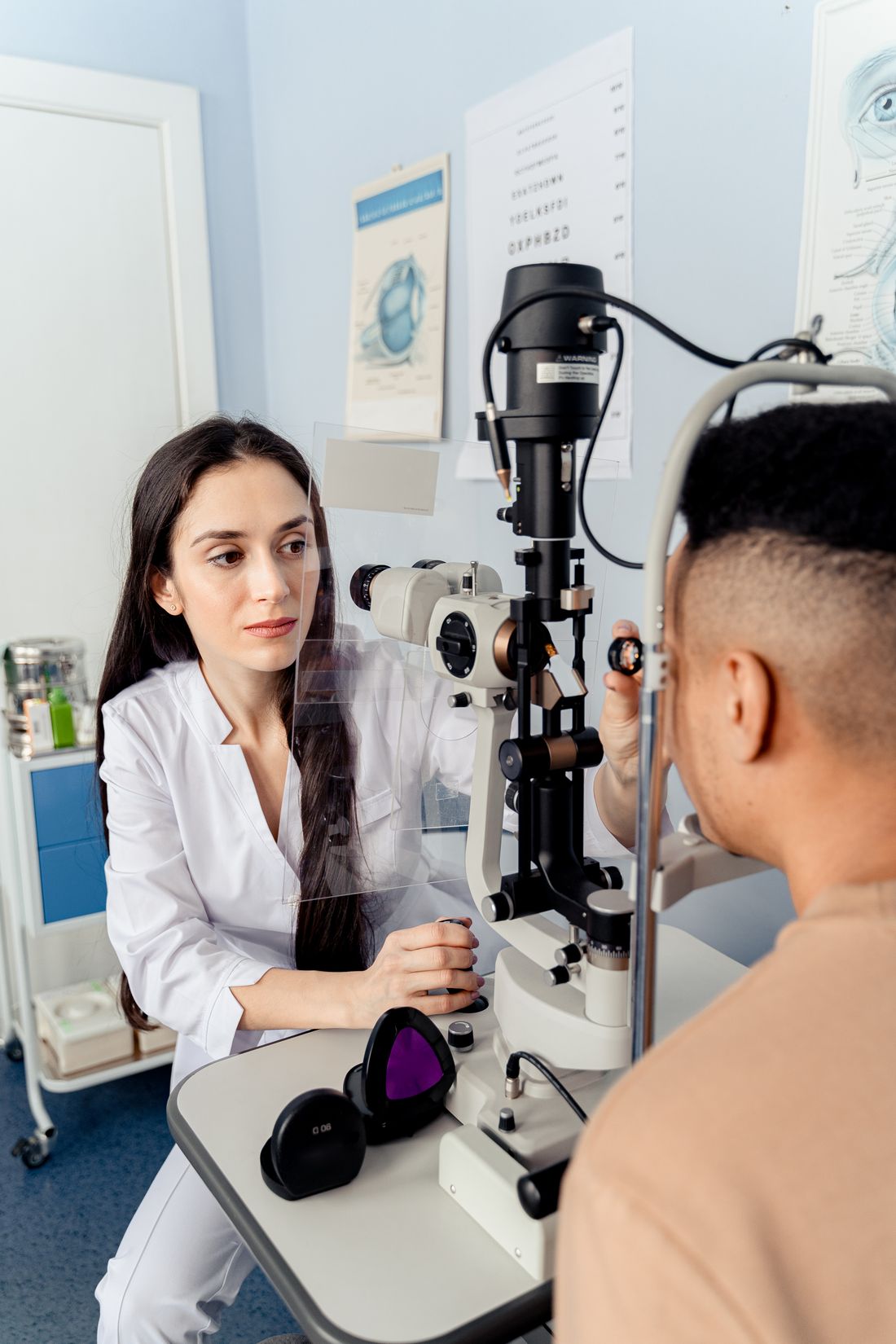
Slit Lamp Examination
Slit-lamp biomicroscopy allows magnified examination of the front of the eye, including the cornea, iris and lens.
Understanding the tools clinicians may use to assess your eyes.
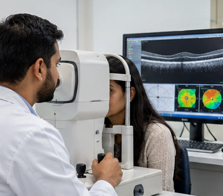
About these images: Genuine documentary/stock photographs and clinical scan images show examples of common eye tests. They do not represent Lensora's staff, premises or equipment. Please confirm available testing before booking.
Slit-lamp biomicroscopy allows magnified examination of the front of the eye, including the cornea, iris and lens.

OCT provides cross-sectional retinal images that can help assess the macula, retinal layers and optic nerve.

A dilated examination, when appropriate, can help evaluate the retina, macula and peripheral retina.

Tonometry measures pressure inside the eye as part of a broader glaucoma evaluation.
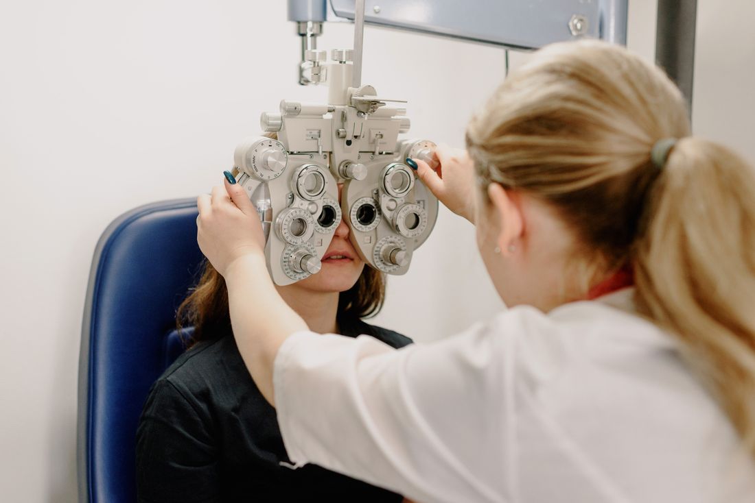
Vision charts and refraction testing help measure sight and determine whether glasses may improve clarity.

Visual-field testing assesses how well light is detected across the field of vision, which can help evaluate optic-nerve conditions.
Understand how an OCT scanner, slit-lamp microscope, tonometer and other common tools are used in eye care.
Explore Modern Equipment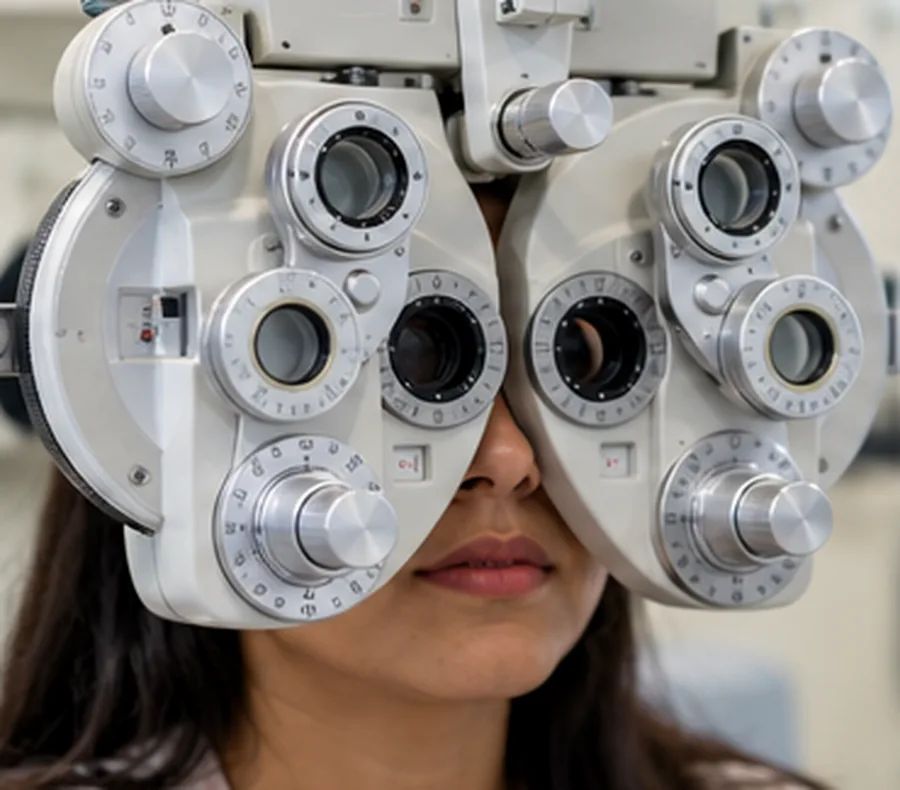
Bring previous eye tests, scan reports, prescriptions and your current medication list. Some examinations use dilating drops that temporarily blur vision. Ask the clinic whether you will need someone to accompany you.
Request an AppointmentTell us what you need, and request a visit with the Lensora team.